Everything below comes from a single subject: me. That is the central limitation and I am not going to bury it. What these findings can do is establish that something physical and visible is happening, in a disease that is routinely described as having no objective findings.
Chest imaging
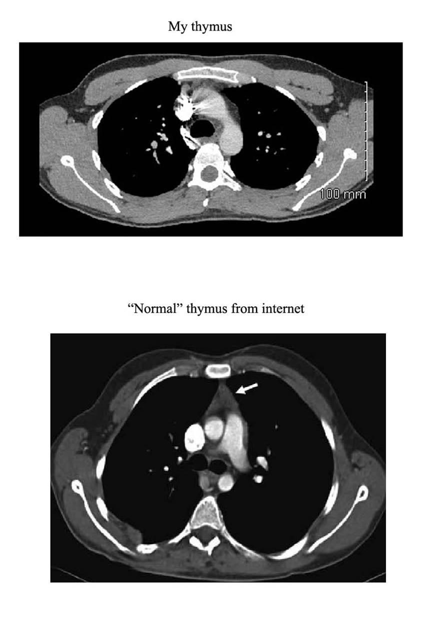
This is the region where I have always felt the root of the illness was, and it is where I would expect to see the effects of epithelial progenitor depletion. It could equally be developmental variation, and I want to be clear that one scan read as normal by a radiologist is weak evidence on its own. What makes me keep returning to it is the anatomy: compression here could impair the thoracic duct and the lymphatics, and this area is densely innervated by the vagus nerve.
It also offers a different reading of the adrenergic autoantibodies that have attracted so much attention in ME/CFS and POTS research. If those antibodies arise from microbial molecules translocating across a permeable gut barrier and cross-reacting with host receptors, then they are a marker of barrier permeability rather than a primary autoimmune driver.
Skin and vasculature
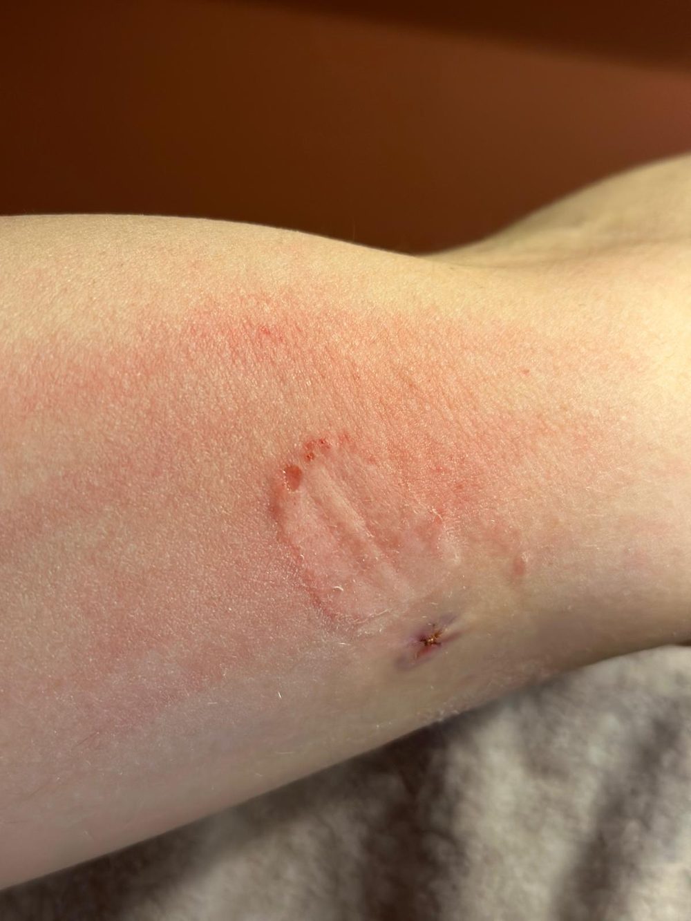
The bandage photograph is the one I show people who tell me this illness is a problem of neural sensitisation. Nothing about a sensitised nervous system removes skin. This is an acquired chemical vulnerability with a visible physical result, from a product I had used my whole life without incident.
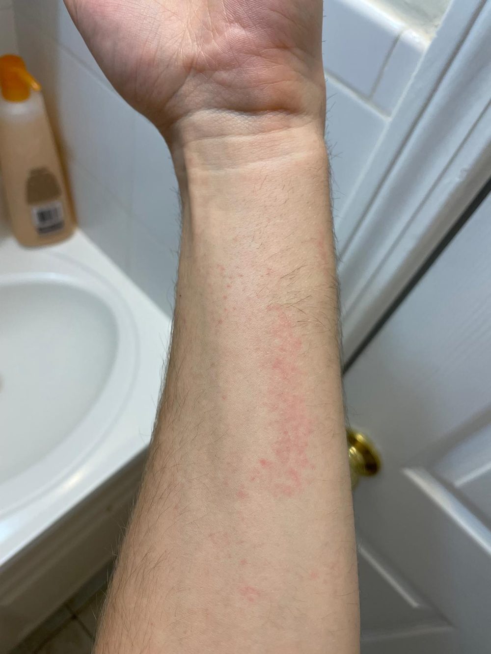
The red dots are not a rash — not raised, not itchy, not allergic. That is blood. Titanium dioxide nanomaterials have been shown to cause endothelial cell leakiness by disrupting VE-cadherin, which is the mechanism I would expect to produce exactly this.
The same exposure produced striations deep in the skin shortly afterwards, which faded over the following year. Titanium dioxide has been shown to cause separation of muscle fibres through disruption of junction proteins.
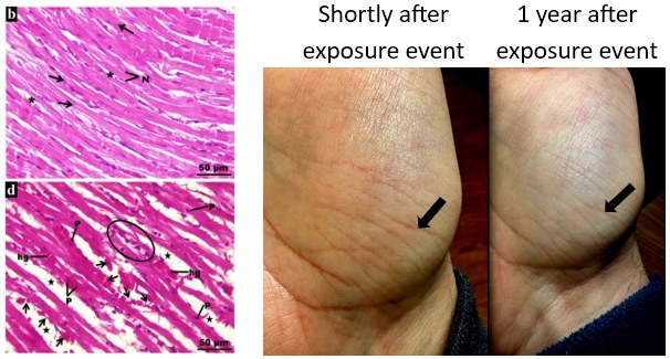
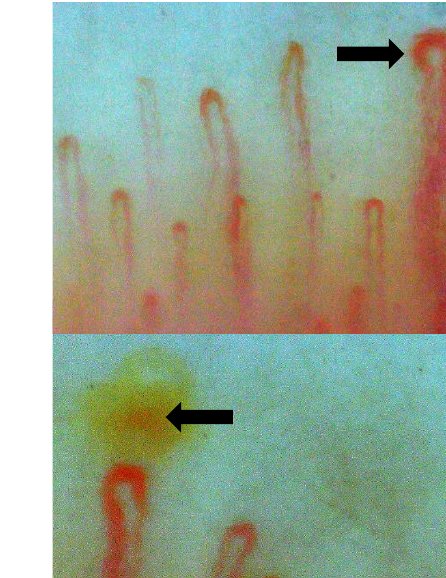
Those dark spots are the finding I find most interesting. They could be haemosiderin from vessel damage, or they could be the microclots themselves. They are slightly larger than the microclots seen in blood testing, which would fit if clots continue to grow before becoming visible in the nailbed.
Amyloid-fibrin microclots have become a major focus in Long COVID and ME/CFS research. My question is whether nanoparticles could be nucleating them. There is published work showing that both albumin and fibrinogen adsorb irreversibly to titanium dioxide surfaces. If a single particle binds multiple fibrinogen molecules, that would offer a route to clot formation without the usual clotting factors.
Electron microscopy
Standard neuropathy testing counts nerve density in a punch biopsy. That test cannot see the kind of damage I was looking for. So in 2023 I had a 3 mm punch biopsy taken 3 cm above the medial side of my left elbow and imaged with transmission electron microscopy.
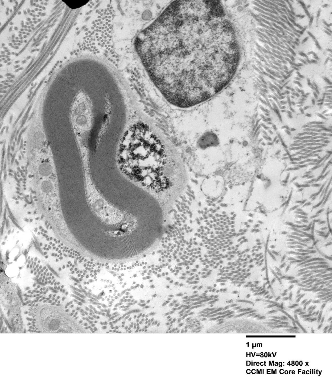
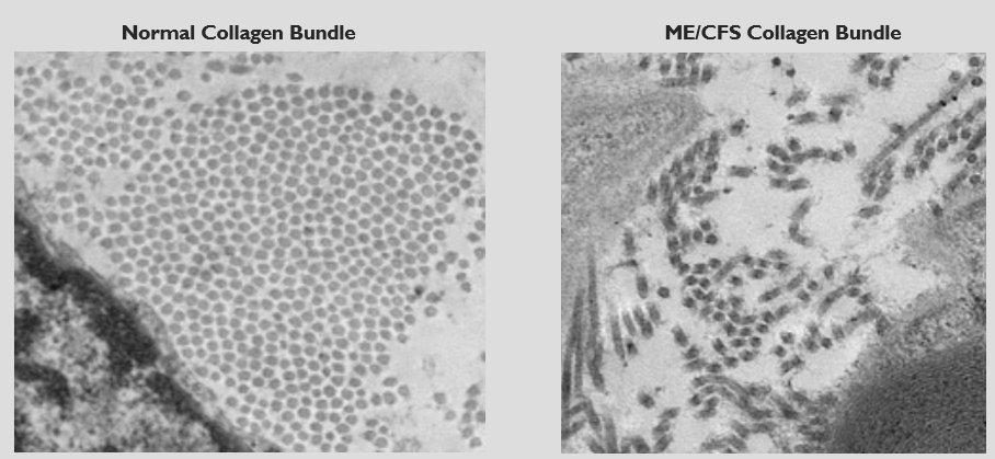
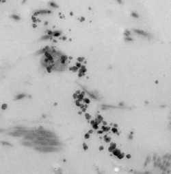
I sent these images to around a dozen nerve morphologists. Two of their assessments:
Three things stand out to me. Glycogen deposits and Nissl substance in mass, pressing on nerves. Pockets of demyelination that do not look like MS or dense degeneration, and look more like puncture or tearing. And glycogen deposits in keratinocytes, the cells that form the epithelial barrier.
On the collagen: fibril diameter and fibril counts were normal, but the bundles were separated and disorganised. A synthesis defect would be expected to change the fibrils themselves. Normal fibrils in disordered bundles reads to me as mechanical disruption after the fact.
The dense particles in the third image are the least settled finding here. Size, shape and composition all affect how a particle interacts with a given cell type, and I have not been able to get compositional analysis done. I am not claiming these are titanium dioxide. I am saying they warrant testing.
The broader point is methodological: a nerve density count would have returned normal on this sample. If TEM shows abnormalities that density counts miss, then neuropathy testing in this patient population needs to change.
Exposure categories
Four classes of exposure, four symptom sets. I can identify which one I am in by which set appears. There is no overlap between them, which is the observation the whole model rests on.
| Exposure type | Symptoms | What sets it apart |
|---|---|---|
| Volatile reactive substances cleaning chemicals, solvents |
|
Stimulated during the exposure, crash after leaving it |
| Abrasive particles titanium dioxide |
|
Progresses tissue depletion. The only exposure where symptoms keep worsening after I leave it |
| Polymers and fibres plastics, rubber, mulch, adhesives |
|
Builds up over time and persists permanently after the exposure ends. Less adhesive material may migrate around the body |
| Mold |
|
Effects are temporary |
Metabolic findings
My organic acid panel came back chronically low across the board — succinic, methylsuccinic, glutaric, malic, adipic, pyruvic, isocitric and citric all below reference range, several at zero. Rats exposed to titanium dioxide show a broad decrease in organic acid levels on NMR spectra that persists chronically. This is the sort of progressive deterioration in energy production that would connect to the metabolic disturbances already described in ME/CFS.
References
These are the published studies the model draws on. They are real and independent of me; the synthesis and the application to ME/CFS are my own.
- Weisz G. Epidemiology and health care reform: the National Health Survey of 1935–1936. Am J Public Health. 2011;101(3):438–47.
- Centers for Disease Control and Prevention. Chronic Diseases in America. 2022.
- Lee PH, Park S, Lee YG, Choi SM, An MH, Jang AS. The impact of environmental pollutants on barrier dysfunction in respiratory disease. Allergy Asthma Immunol Res. 2021;13(6):850–62.
- Deinhardt-Emmer S, et al. SARS-CoV-2 causes severe epithelial inflammation and barrier dysfunction. J Virol. 2021;95(10):e00110-21.
- Sfera A, et al. Endothelial senescence and chronic fatigue syndrome, a COVID-19 based hypothesis. Front Cell Neurosci. 2021;15:673217.
- Rawski DP. Pulp and paper: nonfibrous components. In: Encyclopedia of Materials: Science and Technology. Elsevier; 2001. p. 7908–10.
- Kreyling WG, et al. Quantitative biokinetics over a 28 day period of freshly generated, pristine, 20 nm titanium dioxide nanoparticle aerosols in healthy adult rats. Part Fibre Toxicol. 2019;16:29.
- Warheit DB, et al. Inhalation of high concentrations of low toxicity dusts in rats results in impaired pulmonary clearance mechanisms and persistent inflammation. Toxicol Appl Pharmacol. 1997;145(1):10–22.
- EFSA Panel on Food Additives and Flavourings. Safety assessment of titanium dioxide (E171) as a food additive. EFSA Journal. 2021;19(5):6585.
- Shi H, Magaye R, Castranova V, Zhao J. Titanium dioxide nanoparticles: a review of current toxicological data. Part Fibre Toxicol. 2013;10:15.
- Chen J, Dong X, Zhao J, Tang G. In vivo acute toxicity of titanium dioxide nanoparticles to mice after intraperitoneal injection. J Appl Toxicol. 2009;29(4):330–7.
- Lee YG, Lee SH, Hong J, Lee PH, Jang AS. Titanium dioxide particles modulate epithelial barrier protein, Claudin 7 in asthma. Mol Immunol. 2021;132:209–16.
- Gao Y, Gopee NV, Howard PC, Yu LR. Proteomic analysis of early response lymph node proteins in mice treated with titanium dioxide nanoparticles. J Proteomics. 2011;74(12):2745–59.
- Pérez-Arizti JA, et al. Titanium dioxide nanoparticles promote oxidative stress, autophagy and reduce NLRP3 in primary rat astrocytes. Chem Biol Interact. 2020;317:108966.
- Suker DK, Jasim FA. Liver histopathological alteration after repeated intra-tracheal instillation of titanium dioxide in male rats. Gastroenterol Hepatol Bed Bench. 2018;11(2):159–68.
- El-Bestawy EM, Tolba AM. Effects of titanium dioxide nanoparticles on the myocardium of the adult albino rats and the protective role of β-carotene. J Mol Histol. 2020;51(5):485–501.
- Setyawati MI, et al. Titanium dioxide nanomaterials cause endothelial cell leakiness by disrupting the homophilic interaction of VE-cadherin. Nat Commun. 2013;4:1673.
- Visser VL, Rusinek H, Weickenmeier J. Peak ependymal cell stretch overlaps with the onset locations of periventricular white matter lesions. Sci Rep. 2021;11:21956.
- Joseph P, et al. Insights from invasive cardiopulmonary exercise testing of patients with myalgic encephalomyelitis/chronic fatigue syndrome. Chest. 2021;160(2):642–51.
- Hives L, et al. Can physical assessment techniques aid diagnosis in people with chronic fatigue syndrome/myalgic encephalomyelitis? A diagnostic accuracy study. BMJ Open. 2017;7(11):e017521.
- Huggins CB, Froehlich JP. High concentration of injected titanium dioxide in abdominal lymph nodes. J Exp Med. 1966;124(6):1099–106.
- Valentini X, et al. Hepatic and renal toxicity induced by TiO₂ nanoparticles in rats: a morphological and metabonomic study. J Toxicol. 2019;2019:5767012.
- Korábková E, et al. Behaviour of titanium dioxide particles in artificial body fluids and human blood plasma. Int J Mol Sci. 2021;22(19):10614.