The claim is narrow and specific. It is not that ME/CFS is caused by stress, or by a toxin in the ordinary sense. It is that a damaged epithelial barrier admits material into the body that then causes mechanical injury faster than the body can repair it.
Medicine has largely treated patients as though they exist in a vacuum. We do not. We are in continuous exchange with the air around us, and for someone whose barriers have been compromised, that exchange stops being neutral.
I also want to be precise about a word. I reject dysfunction as a description of this illness and use damage instead. If the problem were a dysfunction — a system operating incorrectly — then out of the hundreds of drugs and supplements patients have tried, you would expect at least one to move the needle. The near-total failure of everything is itself evidence that the body is already compensating as well as it can, around an injury that has not been removed.
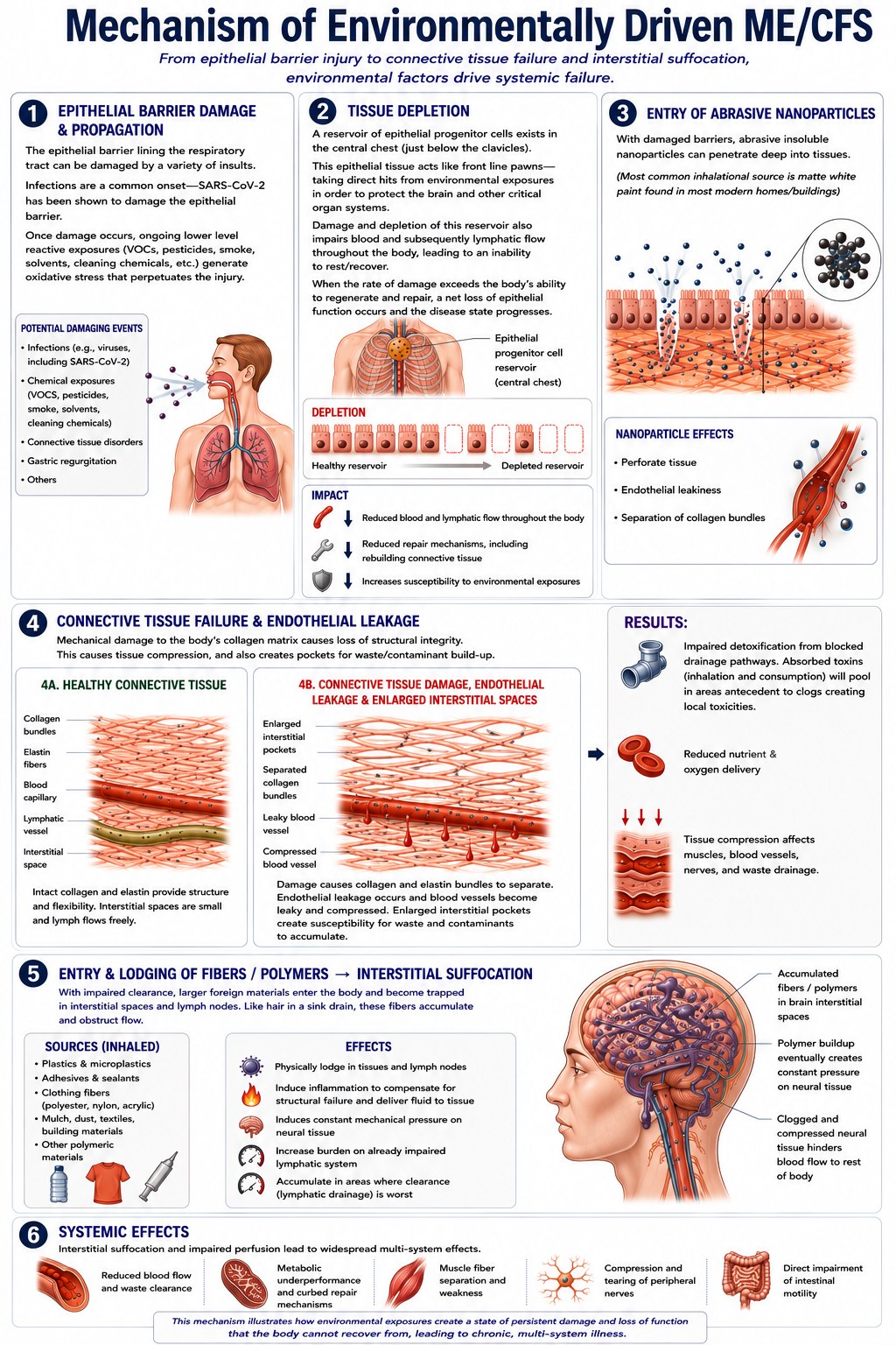
The six stages
-
Epithelial barrier damage and propagation
The epithelial barrier lining the respiratory tract can be damaged by several kinds of insult: infection, including SARS-CoV-2, which has been shown to damage the barrier directly; chemical exposure to VOCs, pesticides, smoke, solvents and cleaning products; connective tissue disorders such as EDS; and gastric regurgitation.
Once damage occurs, ongoing lower-level reactive exposure generates oxidative stress that perpetuates the injury. The result is an acquired environmental susceptibility. This connection is well established for allergies and asthma. It has not been made for ME/CFS or multiple chemical sensitivity.
-
Tissue depletion
There is a reservoir of epithelial progenitor cells in the central chest, just below the clavicles. These cells act like front-line pawns, taking the direct hits from environmental exposure in order to protect the brain and other critical organs.
As that reservoir depletes, blood and lymphatic flow are impaired throughout the body, which impairs the ability to rest and recover. When the rate of damage exceeds the rate of regeneration, there is a net loss of epithelial function and the disease state progresses. Each round leaves fewer buffer cells for the next exposure, which is why this gets worse over time at a constant level of exposure.
-
Entry of abrasive nanoparticles
With the barrier damaged, insoluble nanoparticles can penetrate deep into tissue. Titanium dioxide is the one I focus on: chemically inert, extremely hard, used as an abrasive in polishing compounds and in toothpaste, cleared from the body very slowly, and absorbed rapidly when inhaled.
The most common inhalational source is matte white paint, which is high in titanium dioxide and low in binder, so the surface sheds. This is usually described as a rare industrial exposure. It is not rare. It is on the walls of most modern homes and buildings. What is rare is an exposure high enough to cause acute symptoms in a healthy person.
These particles perforate tissue, make blood vessel walls leaky, and separate collagen bundles.
-
Connective tissue failure and endothelial leakage
Mechanical damage to the collagen matrix costs the tissue its structural integrity. In healthy connective tissue, intact collagen and elastin provide structure and flexibility, interstitial spaces are small, and lymph flows freely.
Once collagen and elastin bundles separate, interstitial pockets enlarge and become places for waste and contaminants to collect. Blood vessels become leaky and compressed. Detoxification is impaired because drainage pathways are blocked — absorbed toxins pool in the areas upstream of the blockage and create local toxicities. Nutrient and oxygen delivery drops. Tissue compression affects muscles, vessels, nerves and waste drainage all at once.
-
Entry and lodging of fibres and polymers
With clearance already impaired, larger foreign materials — plastics and microplastics, adhesives and sealants, clothing fibres such as polyester, nylon and acrylic, mulch, dust and building materials — enter and become trapped in interstitial spaces and lymph nodes. Like hair in a sink drain, they accumulate and obstruct flow.
They lodge physically in tissue, induce inflammation as the body attempts to compensate for the structural failure, exert constant mechanical pressure on neural tissue, and gather where lymphatic drainage is worst. I call the result interstitial suffocation, because that is what it feels like.
-
Systemic effects
Impaired perfusion and interstitial suffocation produce effects across every system: reduced blood flow and waste clearance, metabolic underperformance and curbed repair, muscle fibre separation and weakness, compression and tearing of peripheral nerves, and direct impairment of intestinal motility.
This is where the model connects to what is already documented in ME/CFS — impaired peripheral oxygen extraction, lymphatic congestion, white matter hyperintensities, low organic acids, microclots — without needing a separate explanation for each.
Exposure order matters
The four categories of exposure I have identified do not simply add up. Each one changes how the body responds to the next, which means the same factors arriving in a different order would not produce the same outcome. This is the part of the model I would most want a researcher to take seriously, because it explains why patient presentations vary so widely without requiring them to have different diseases.
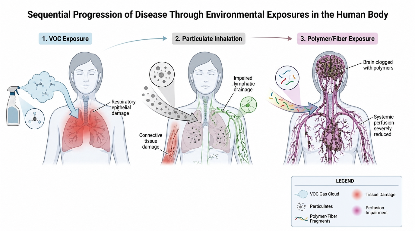
The symptom sets are distinct enough that I can tell which class of exposure I am in by which one appears — see the exposure table. That reproducibility is the strongest argument I have against a sensitisation explanation. If this were a sensitisation or mast cell problem, the reaction would be broadly the same regardless of what triggered it. Mine are not similar at all. They have no overlap, and each one makes sense in the context of what I was exposed to.
What this model would explain
- Why tests come back normal. Titanium dioxide has been shown to induce a chronic fatigue syndrome-like illness in animals at lower doses without producing major structural or blood-based biomarker changes.
- Why chemical sensitivity and ME/CFS overlap so heavily. Around 80% of people with ME/CFS in my surveys self-report chemical sensitivities. Given that I had no idea I had environmental issues for four years, I suspect the true figure is higher.
- Why the diagnostic labels blur. On this model, MCS, POTS, ME/CFS, Long COVID, fibromyalgia and EDS are versions of the same problem, differing by genetics, exposure type, and how far the disease state has progressed. Duration may be why we see biomarker differences between Long COVID and ME/CFS cohorts.
- Why mast cell treatment disappoints. Mast cell activation is downstream of barrier permeability, not upstream of it. Treating it does not address what opened the barrier.
- Why pacing works. There is a constant tug of war between healing and damage. Limiting exertion shifts the balance slightly toward healing. It does not resolve the underlying injury.
Open questions
I would rather state these than have them found for me.
- Are the nanoparticles damaging tissue directly through abrasion, or indirectly through MMP-mediated degradation that leaves tissue vulnerable to ordinary mechanical stress? Possibly both. I have not been able to distinguish them.
- How much of what I observe is specific to my own exposure history, and how much generalises? I have argued it generalises, but my sample is one person plus survey data, and survey data is weak evidence.
- Where are the collagen-related SNPs, if connective tissue susceptibility is part of the picture?
- Is the degeneration I describe too rapid to be explained by the mechanism as stated?
- Could viral persistence account for the same observations? I think the evidence there is weak — Paxlovid trials failed, a majority of Long COVID patients show no viral persistence, spike protein levels do not track severity, and healthy controls carry spike too. But it has not been ruled out.
Status of this model. This is a hypothesis developed by a patient with a research background, based on personal observation, test results, and published literature. It has not been peer-reviewed or tested in a controlled setting. The published studies cited on the evidence page are real; the synthesis and the application to ME/CFS are mine, and are unproven.