
This is my model for how environmental factors can drive chronic illness — even in people whose onset was infectious, or otherwise not environmental — and for why this patient population is so heterogeneous. If it is right, a great deal of what looks like dysfunction in ME/CFS is better described as damage: a damaged system has to be protected long enough to repair, and cannot be while the exposure continues. It begins with the epithelial barrier that normally keeps airborne contaminants out of the body. Once that is compromised, ordinary indoor air becomes a source of continuous injury, and the damage outpaces repair.
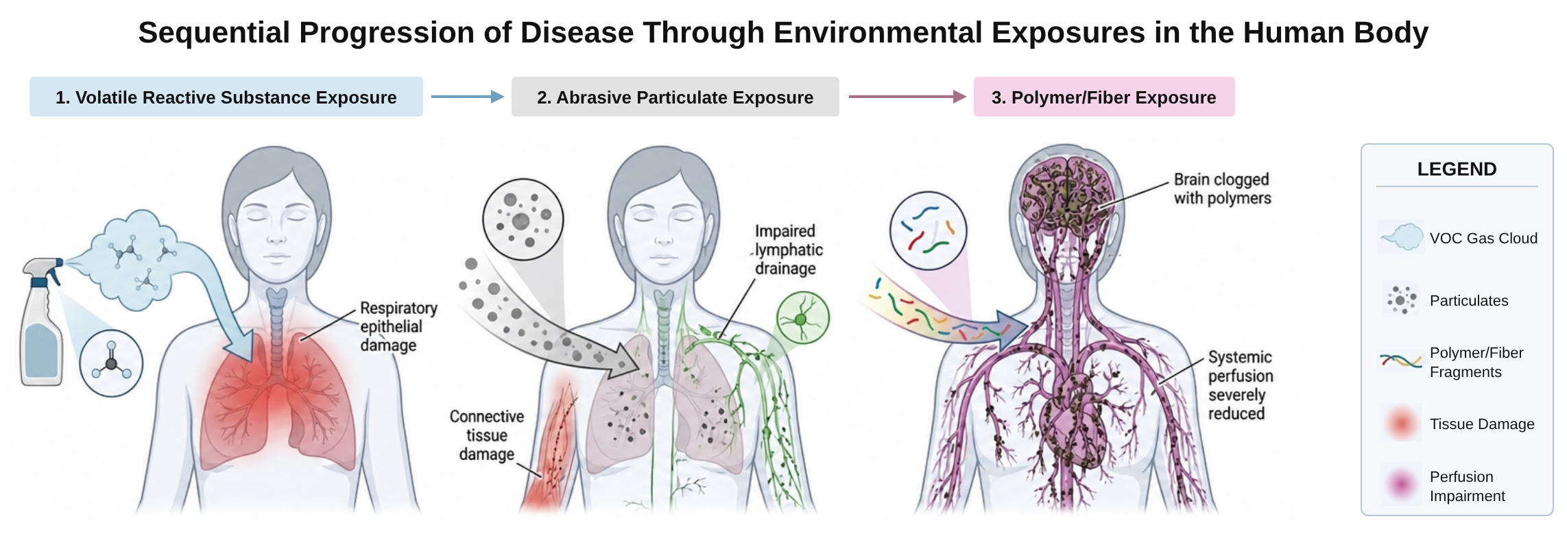
The mechanism runs in stages:
- A damaged epithelial barrier admits abrasive nanoparticles that perforate tissue and pry apart the fibers within collagen bundles
- Producing connective tissue failure and endothelial leakage
- Fibers and polymers then lodge in the resulting interstitial spaces
- Producing interstitial suffocation and impaired perfusion throughout the body.
Medicine has largely treated patients as though they exist in a vacuum. We do not. We are in continuous exchange with the air around us, and for someone whose barriers have been compromised, that exchange stops being neutral.
I also want to be precise about a word. I reject dysfunction as a description of this illness and use damage instead. If the problem were a dysfunction — a system operating incorrectly — then out of the hundreds of drugs and supplements patients have tried, you would expect at least one to move the needle. The near-total failure of everything is itself evidence that the body is already compensating as well as it can, around an injury that has not been removed.
When a cell is damaged, the body has three ways to respond, and which one dominates in a given person determines what disease results (M1). A hyperproliferative response — unregulated replication to replace the damaged cells — can produce cancer. A hyperactive immune response, clearing cells the body judges to be damaged, can produce autoimmune disease. A hypoproliferative response — no compensatory growth, and no immune clearance either — produces neither. It produces cell death and loss of tissue and function on their own. I think that is what multiple chemical sensitivity and ME/CFS are: a hypoproliferative response to the same underlying damage that produces cancer or autoimmunity in other people.
That hypoproliferative response is what the mechanism below traces in full, stage by stage.
For evidence that an environmental explanation is plausible at a population level — independent of my own case — see the Context page: how much more common chronic illness has become, what's changed in the chemical and material environment, and precedent from other diseases that turned out to have environmental drivers.
The five stages
-
Epithelial barrier damage
The epithelial barrier lining the respiratory tract can be damaged by several kinds of insult (M3). Infection is a common onset; SARS-CoV-2 has been shown to damage the barrier directly.
What starts it
InfectionViruses, including SARS-CoV-2Chemical exposureVOCs, pesticides, smoke, solvents, cleaning productsConnective tissue disordersSuch as EDSGastric regurgitationReflux reaching the airwayOther insultsFurther triggers not listed hereWhat keeps it going
Once the barrier is damaged, ongoing lower-level exposures to reactive chemicals (VOCs, pesticides, smoke, solvents, cleaning products) generate oxidative stress that keeps the injury going.
M3 — An intact epithelial barrier, and the same barrier with a cell lost from it. Once damage occurs, ongoing lower-level reactive exposure generates oxidative stress that perpetuates the injury. -
Tissue depletion
Once damage occurs, ongoing lower-level reactive exposure generates oxidative stress that perpetuates the injury. The result is an acquired environmental susceptibility. This connection is well established for allergies and asthma. It has not been made for ME/CFS or multiple chemical sensitivity.
The airways are lined by an epithelium that is constantly maintained by its own stem cells. Basal cells sit along the base of the lining and divide to replace damaged or lost cells, and the glands in the walls of the larger airways hold additional progenitors that help after more severe injury (M2B A). These progenitors are most abundant in the trachea and main bronchi, the central airways that take the heaviest share of everything we breathe in. Every time inhaled exposures kill lining cells, progenitors are spent replacing them.
My hypothesis is that repeated injury eventually uses up that repair capacity faster than it can recover, leaving the barrier thinner, slower to heal and easier to breach, and that this depletion is concentrated in the central chest, where exposure is greatest (M2B B).
I think the loss may not stop at the lining. Repeated epithelial loss, and the chronic, low-grade inflammation that goes with it, could over time also reduce the soft tissue around the central airways. One place that soft tissue can be seen is the retrosternal space, between the back of the sternum and the great vessels, which in adults normally holds what remains of the thymus mixed with fat, along with small vessels, lymph nodes and connective tissue. On my own 2017 chest CT, that space holds noticeably less soft tissue than on a typical scan (E7 on the Evidence page). The radiologist read the scan as normal, meaning no mass or enlarged lymph nodes were seen; the reduced bulk is a qualitative observation rather than a measurement. It is consistent with the depletion model, but it doesn't prove it on its own. The thymus also normally shrinks and is replaced by fat in adulthood, so a smaller retrosternal space can be part of normal aging, which is another reason I treat this as a hypothesis rather than a finding.
With less tissue in that space, the great vessels and the structures behind them sit closer to the back of the sternum, with less tissue cushioning them from the chest wall. That could amplify both mechanical and chemical effects there, and it fits two things I noticed: the "crack" I could feel forming in my chest, and being able to turn symptoms on and off by tightening my chest muscles (both described in Tissue depletion on the Evidence page).
As repair capacity runs down, the damage doesn't stay local. The central chest, where my own scan shows that reduced soft tissue, is also where the body's circulation converges, so damaged, inflamed and depleted tissue there could reduce blood flow everywhere (M2B C). In my model, it could do this in three ways. First, every drop of blood bound for the body passes through the lungs before it reaches the left side of the heart. Inflamed, damaged tissue in the chest raises the resistance in that circuit, so less blood reaches the left heart and less is pumped out to the rest of the body. Second, breathing is itself a pump: each breath lowers the pressure in the chest and draws venous blood and lymph back toward the heart. A damaged, guarded chest breathes shallowly, and that pump weakens. Third, lymph from most of the body drains back into the bloodstream just below the left collarbone. Swelling and congestion there slow drainage from everywhere upstream, and fluid that can't drain is fluid that isn't circulating.
The result is reduced blood flow and oxygen delivery throughout the body, which impairs the ability to rest and recover. When the rate of damage exceeds the rate of regeneration, there is a net loss of epithelial function and the disease state progresses. Each round leaves fewer buffer cells for the next exposure, which is why this gets worse over time at a constant level of exposure (M4).

Illustration M2B — Tissue depletion. (A) Established airway anatomy: ciliated and goblet cells line the airway on a single row of basal cells, the lining's stem cells, and submucosal glands hold extra progenitors in the larger airways; basal cells are most numerous in the trachea and main bronchi. The magnified view shows a basal cell dividing and its daughter cell moving up to fill a gap in the lining. (B) and (C) show the proposed model. (B) Repeated injury uses up the basal cells until lining cells that are lost are no longer replaced; the note beside it proposes that the same process may also leave less soft tissue in the retrosternal space. (C) Two routes by which damage in the chest could reduce blood flow to the whole body, and the narrowed retrosternal compartment (reduced thymic remnant + mediastinal fat) in front of the great vessels. Illustrations adapted from Servier Medical Art (CC BY 4.0). Step 0 of 3The Threshold Model
How ambient VOC/toxic exposures in the home can perpetuates and progress tissue depletion following an initial damaging event, such as from an infection or other insult.
VOC Exposure Remains Constant For Each 24 Hour Simulated CycleAirway1Cycle12VOC molecules12Cells remaining1 : 1VOC : cellwithin capacityA healthy barrier is able to buffer against ambient VOC exposures in the home.Larger insults like infections, pollution, and connective tissue damage will damage or destroy part of the barrier.In this example, a viral infection destroys one of our cellsAnd particulate matter from smog damages another cell, causing it to lose its detoxification ability.The cells are able to detoxify all of the VOCs and survive.The toxic load per cell has now risen without the exposure increasing.Damaged cells then lose part of their capacity to buffer against toxic exposures, which makes them even more susceptible to future exposuresWhen the damage becomes too much, the cells will eventually die.As cells die, the relative exposure size for the remaining cells increases again, even though the total exposure level the person is exposed to remains the same.This process repeats itself until the tissue depletes to nothing, with even smaller exposure causing more damage as the relative exposure size skyrockets.
Note how the VOC:cell ratio is increasing, which is why the cells are becoming damaged from the same exposure.
The relative exposure for these cells increased as the absolute number of buffering cells declined from the prior insult.
Healthy epithelial barrier cellsNon-Damaging Cycle
Large Cellular Insult
First Damaging Cycle
First Cycle at Which Cells Begin to Die from Ambient Exposures
Damage Progresses
Damage and Depletion Accelerates
Depletion Has Concluded
Release an exposureStart from cycleSpeedM4 — The Threshold Model. -
Entry of abrasive nanoparticles and connective tissue failure
Illustration M2C — How insoluble particles reach tissue once the airway barrier is damaged. A: three effects reported for titanium dioxide nanoparticles: tight-junction disruption, endothelial leakiness, and (my hypothesis) connective tissue damage. B: the same airborne load meets an intact barrier and a damaged one; only through the damaged barrier do particles reach the tissue beneath. Under the intact barrier, intact collagen and elastin provide structure and flexibility and lymph flows freely; under the damaged one, the fibers in those bundles have separated, interstitial pockets have enlarged, and blood vessels are leaky and compressed. C: consequences of connective tissue failure: lymphatics pinched before the lymph node so absorbed toxins back up; muscle changes reported in both ME/CFS and Long COVID (thickened capillary basement membranes, abnormal subsarcolemmal mitochondria, lower oxygen uptake after exertion); and collapse of disorganized, mechanically fragile tissue onto muscles, vessels, nerves and drainage routes. Particles are drawn far larger than life. Sources for A: Brun et al., 2014; Smallcombe et al., 2020; Setyawati et al., 2013. Sources for the muscle findings in C: Scheibenbogen & Wirth, 2024; Bizjak et al., 2024; Aschman et al., 2023; Slaghekke et al., 2025. Magnified inset: commercial titanium dioxide nanopowder under transmission electron microscopy, scale bar 50 nm. Fresegna et al., Nanomaterials, 2021, CC BY 4.0. With the barrier damaged, insoluble nanoparticles can penetrate deep into tissue (M2C). Titanium dioxide is the one I focus on: chemically inert, extremely hard, used as an abrasive in polishing compounds and in toothpaste, and absorbed rapidly when inhaled. Reported half-life figures are commonly around 60–70 days, but the half-life is dose-dependent — a minute exposure clears relatively quickly, while a large exposure event can push clearance from days out toward years. The same reasoning applies to other relatively inert, insoluble nanoparticles — silica, aluminum oxide, calcium carbonate — though titanium dioxide is the one I have focused on and have the most evidence for. The EU has already removed it from the approved list of food additives, after its food safety agency could not rule out a risk of genotoxicity and could not establish a safe daily intake — the same substance remains in widespread use in the United States, including in food.
The most common inhalational source is matte white paint, which is high in titanium dioxide and low in binder, so the surface sheds. This is usually described as a rare industrial exposure. It is not rare. It is on the walls of most modern homes and buildings. What is rare is an exposure high enough to cause acute symptoms in a healthy person.
Picture a piece of construction paper coated in glue stick with a handful of sand thrown on it. Come back a year later and roughly half the sand has fallen off. That is what is happening at a microscopic and nanoscopic scale on a wall painted in matte white: tiny particles work loose from the surface into the air. Larger microparticles settle to the ground within a few hours, but these nanoparticles are small enough that they never settle — once they are in a room's air, they simply keep circulating.
These particles perforate tissue, make blood vessel walls leaky, and pry apart the fibers inside collagen bundles.
Mechanical damage to the collagen matrix costs the tissue its structural integrity. In healthy connective tissue, intact collagen and elastin provide structure and flexibility, interstitial spaces are small, and lymph flows freely.
Once the fibers in collagen and elastin bundles separate and lose alignment, interstitial pockets enlarge and become places for waste and contaminants to collect. Blood vessels become leaky and compressed. Detoxification is impaired because drainage pathways are blocked — absorbed toxins pool in the areas upstream of the blockage and create local toxicities. Nutrient and oxygen delivery drops. Tissue compression affects muscles, vessels, nerves and waste drainage all at once. This is also my working explanation for the joint hypermobility described on the Personal Journey page — ligament laxity from structural damage to connective tissue, not a separate connective tissue disorder.
I don't think autoimmunity or MMP-driven breakdown is doing much of this — I think it's mostly impaired collagen production, on top of the mechanical disruption described above. A barrier that's spending its resources on damage control isn't well placed to keep resynthesizing the structural proteins it depends on.
-
Entry and Lodging of Fibers and Polymers

Panel A images (scanning electron micrographs, cropped): 1 Jemec Kokalj A. et al., Polymers 2021, CC BY 4.0; 2 Li N. et al., Polymers 2025, CC BY 4.0; 3 Kärkkäinen N. and Sillanpää M., Environmental Science and Pollution Research 2021, CC BY 4.0; 4 Zhang H. et al., Molecules 2026, CC BY 4.0; 5 Moorhead A. and Francis L.F., Journal of Coatings Technology and Research 2025, CC BY 4.0; 6 Lin P. et al., Materials & Design 2021, CC BY 4.0.
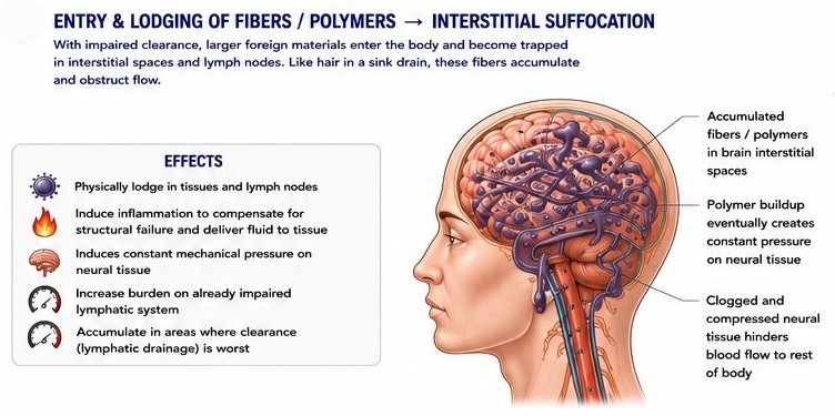
M2D — Larger foreign materials enter and become trapped in interstitial spaces and lymph nodes, where they lodge physically in tissue, induce inflammation, and exert constant mechanical pressure on neural tissue. With connective tissue damage and tissue depletion already impairing blood flow and waste clearance, larger foreign materials — including, but not limited to, plastics and rubber, adhesives and sealants, clothing fibers such as polyester, nylon and acrylic, mulch, sawdust and paint — enter and become trapped in interstitial spaces and lymph nodes (M2D). Like hair in a sink drain, they accumulate and obstruct flow. Clogging by these fibers is close to inevitable. It's like a toilet with weak water pressure: sooner or later, it clogs. Poor blood flow leads to the same problem, especially when things that shouldn't go down the drain, like paint and glue, are going down it anyway. You already know those substances are guaranteed to cause a clog in a pipe. In a human body with barrier damage and poor blood flow, why would we expect anything different?
These substances lodge physically in tissue and help drive inflammation as the body tries to compensate for the structural failure. That inflammation ends up exerting constant mechanical pressure on neural tissue and accelerating the damage. I call the result interstitial suffocation, because that is what is happening: these polymeric substances are physically blocking oxygen from reaching the brain.
Think of the blood vessels as a tap delivering fluid to the tissues, and the spaces between cells are the sink where that fluid pools. The lymphatic system is the drain that carries away the excess fluid and cellular waste. Like any drain, it can clog.
By definition, these exposures don't clear well once they're inside the body: they lodge in narrow spaces and build up over time, like hair collecting in a drain. In the brain, that build-up would hinder perfusion and cellular waste drainage, which would cause local toxicities. Through autonomic, inflammatory, and chemosensory means, the body can sense that impaired perfusion and attempts to compensate for it by sympathetic/inflammatory processes to drive blood to the brain tissue. Of course, this attempt fails because things like industrial grade adhesives will easily resist small increases in pressure. The result is that this just raises intracranial pressure and increases damage/pressure on the brain leading to brain fog so many patients describe. Elsewhere in the body, these same clog-prone substances can also accumulate in places other than the brain, which contributes to systemic symptoms. However, if you take an anatomical view of the human body, virtually everywhere except the brain is subjected to relatively strong mechanical forces from muscle tension, to restrictive clothing, and the largest being gravitational forces from the person's own body weight (particularly when lying down). Because the brain is protected by the skull, mechanical forces driving lymphatic flow are almost entirely dependent upon the vasculature system. This makes this waste clearance system particularly vulnerable to cardiac and endothelial insults.
A perspective published in October 2026 by Nemat-Gorgani, Jensen and Davis proposes that impaired waste clearance from the brain may contribute to both ME/CFS and Long COVID (Nemat-Gorgani et al., 2026). They describe the glymphatic and meningeal lymphatic systems as one integrated route. Cerebrospinal fluid flushes waste out of brain tissue and drains through the meningeal lymphatics toward the lymph nodes of the neck, mostly during deep sleep. Autophagy, each cell's internal recycling system, works alongside that flow and is also most active during sleep. When aggregates overload astrocytes and clog the spaces around blood vessels, the aquaporin-4 water channels that drive glymphatic flow can become mislocalized, and clearance slows further. The authors focus on misfolded, amyloid-like proteins rather than plastics, but they list chemicals, heavy metals, toxins and pesticides among the factors that may impair autophagy. Their framework describes the same drainage route as the model here, and the same trap: material that can't be cleared obstructs the pathways that would clear it. Synthetic polymers and insoluble particles add a load that autophagy did not evolve to break down, which is why I think they belong on that list.
-
Tissue and brain die-off
Because of mechanical pressure and reduced perfusion, even subtle neurological demand becomes equivalent to excitotoxicity, given the energy and resources available to the tissue. Tissue in the chest dies off first, and parts of the brain die off in later stages (M6).
The brain die-off is not whole regions dying at once, as in Parkinson's disease, but scattered individual cells (M5). Remaining cells can compensate for the function lost from those cells, but doing so requires even more energy. That added demand produces extreme weakness and fatigue, and leaves the compensating cells susceptible to the same excitotoxic die-off in turn.
- Plastic particles land and lodge in the tissue
- Collagen fibers stretch and distort under strain
- Neurons die, one at a time
M5 — Not whole regions dying at once, but scattered individual cells. Drag to rotate the network; the model runs until one in seven of the cells is lost. M6 labels
Auditory pathway (amber)
Cochlear nuclei (dorsal and ventral): the brain's first stop for sound from the inner ear; begin sorting timing, pitch and loudness.Superior olivary complex: compares the sound reaching each ear to locate where it comes from; also controls the protective ear reflex and feedback to the inner ear.Inferior colliculus: midbrain hub that combines auditory signals and drives reflex turning toward and startle at sounds.Medial geniculate body: thalamic relay that passes sound to the auditory cortex, and to the amygdala for emotional responses.Primary auditory cortex: conscious hearing (pitch, loudness, speech).Autonomic and breathing centers (red)
Nucleus of the solitary tract: main sensory hub for signals from the body (blood pressure, blood oxygen and CO₂, lung stretch, gut) arriving through the vagus nerve.Dorsal motor nucleus of the vagus: parasympathetic output to the gut and lungs (digestion, gut movement, airway tone).Nucleus ambiguus: controls swallowing and voice, and is the main vagal brake on heart rate.Rostral ventrolateral medulla: main sympathetic drive that sets blood-vessel tone and blood pressure.pre-Bötzinger complex / ventral respiratory group: generates the rhythm of breathing.Retrotrapezoid nucleus (CO₂ sensor): senses CO₂ and drives breathing, especially during sleep.Pontine respiratory group: shapes the breathing pattern and the switch between breathing in and out.Locus coeruleus: main source of noradrenaline in the brain; controls arousal, alertness and sympathetic tone.Area postrema: samples the blood directly (it lies outside the blood–brain barrier) and triggers nausea and vomiting.Relay stations (gray)
Caudal pontine reticular nucleus: drives the startle reflex.Periaqueductal gray: coordinates defense responses, including blood pressure and breathing changes.Amygdala: attaches emotional meaning and fear to sounds.Hypothalamus: master controller of autonomic and hormonal responses.Auditory–motor links (teal)
Primary motor cortexPremotor cortexSupplementary motor areaBroca’s areaPutamenCerebellumSuperior colliculusPontine reticular nucleusArcuate fasciculusM6 — Transparent brain showing the auditory pathway (amber) and the brainstem autonomic and breathing centers (red). The cochlear nuclei sit at the junction of the pons and medulla, a few millimeters from the nucleus of the solitary tract, the vagal motor nuclei, the respiratory groups that set breathing rhythm, and the retrotrapezoid nucleus that senses CO₂ and drives breathing during sleep. Scattered cell loss in this small region could plausibly affect hearing, heart rate, blood-vessel tone and breathing drive together. Schematic: positions approximate, not to scale. Use Cutaway to remove the left half and see the brainstem against the inner surface of the right half. Brain surface adapted from BodyParts3D, © The Database Center for Life Science, licensed under CC Attribution-Share Alike 2.1 Japan. The auditory relay itself does not connect directly to these centers; they are linked indirectly through the reticular formation (startle), the periaqueductal gray, and the amygdala and hypothalamus (shown with Connections on). Teal: the auditory system's links to movement. The arcuate fasciculus carries sound from auditory cortex to premotor and motor areas, the route used to repeat a heard word and correct one's own speech (Hickok & Poeppel, 2007). Auditory cortex also works with premotor cortex, the supplementary motor area, the putamen and the cerebellum to time movement to a beat (Zatorre et al., 2007). In the brainstem, a sudden sound reaches the pontine reticular nucleus and triggers the startle reflex within milliseconds (Koch, 1999), and the inferior colliculus drives the superior colliculus to turn the head and eyes toward a sound.

Systemic effects
Impaired perfusion and interstitial suffocation produce effects across every system: reduced blood flow and waste clearance, metabolic underperformance and curbed repair, muscle fiber separation and weakness, compression and tearing of peripheral nerves, and direct impairment of intestinal motility (M2E).
This is where the model connects to what is already documented in ME/CFS — impaired peripheral oxygen extraction, lymphatic congestion, white matter hyperintensities, low organic acids, microclots — without needing a separate explanation for each.
Panel 1 images: nailfold capillaroscopy video stills from Ronald W. Davis, 'ME/CFS Research', Albert Einstein College of Medicine, 2019 (Open Medicine Foundation, YouTube, at 38:44). Davis reported slower red-cell flow in the ME/CFS patient's capillaries than in the healthy control. Panel 3 images: Appelman B. et al., 'Muscle abnormalities worsen after post-exertional malaise in long COVID', Nature Communications 15, 17 (2024), Fig. 5A–B, cropped; CC BY 4.0. Panel 2 images: Bizjak D.A. et al., 'Functional and Morphological Differences of Muscle Mitochondria in Chronic Fatigue Syndrome and Post-COVID Syndrome', International Journal of Molecular Sciences 25(3), 1675 (2024), Fig. 3A and 3D, cropped; CC BY 4.0. Panel 4 images: Azcue N. et al., 'Small fiber neuropathy in the post-COVID condition and Myalgic Encephalomyelitis/Chronic Fatigue Syndrome: Clinical significance and diagnostic challenges', European Journal of Neurology 32(2), e70016 (2025), Fig. 2, cropped; CC BY. Panel 5 graphs: Chatterton B.E. and Burnet R.B., 'Gastric emptying is slow in chronic fatigue syndrome', BMC Gastroenterology 4, 32 (2004), Figs. 2 and 3; CC BY 2.0.
The plainest way I can summarize this: it is full-body silicosis without fibrosis. ME/CFS, on this model, is an anti-fibrotic disease — the body absorbs this kind of mechanical damage without ever mounting the scarring response that would show up on a standard test.
Exposure order matters
The four main categories of exposures I have identified have unique effects. When a person is exposed to multiple types of exposure classes, each one changes how the body responds to the next. This means that the same factors arriving in a different order would not produce the same outcome (M7). This part of the model also continues to the heterogeneity seen in Complex Chronic Illnesses, but helps us to target specific exposure classes to have the largest possible impact on patient health.
Exposure categories
Four classes of exposure, four symptom sets. I can identify which one I am in by which set appears. There is no overlap between them, which is the observation the whole model rests on.
The symptom sets are distinct enough that I can tell which class of exposure I am in by which one appears. That reproducibility is the strongest argument I have against a sensitization explanation. If this were a sensitization or mast cell problem, the reaction would be broadly the same regardless of what triggered it. Mine are not similar at all. They have no overlap, and each one makes sense in the context of what I was exposed to.
Volatile reactive substances
Cleaning chemicals, solvents, paint, varnish, off-gassing plastics

What I feel
- Stimulated feeling while exposed — eyes wide, short of breath, like being on a drug but not pleasantly
- Chest feels compressed or deflated
- No sensation of rest or relaxation, however long I lie down
Why it looks like this
The tell: stimulated during the exposure, crash on leaving it. The nervous system is overworking to compensate for the damage; when you step out, it crashes.
My experience
In the bedroom I had a completely different set of symptoms, and the mask made no difference to them. I concluded the bedroom had a VOC problem from the wood varnish — molecules small enough to pass straight through the filter.
Drives stage 1 — barrier damageAbrasive insoluble nanoparticles
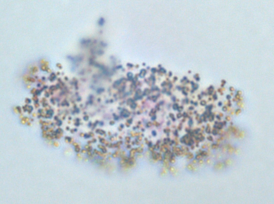
Titanium dioxide, silica, aluminum oxide, calcium carbonate — chiefly matte white paint
What I feel
- Fullness or pressure throughout the body
- Increased pain leading to weakness
- Feeling of cement in the veins
Why it looks like this
The tell: the only exposure where symptoms keep worsening after I leave it. Progresses tissue depletion. These particles are too small to settle out of air and clear from the body slowly.
My experience
I spent four continuous days in a room finished in matte white paint. My partner was in the same room the whole time and was fine. This was the only exposure I have had where the symptoms kept getting worse after I left it.
Drives stage 3 — particle entry, tissue failurePolymers and fibers
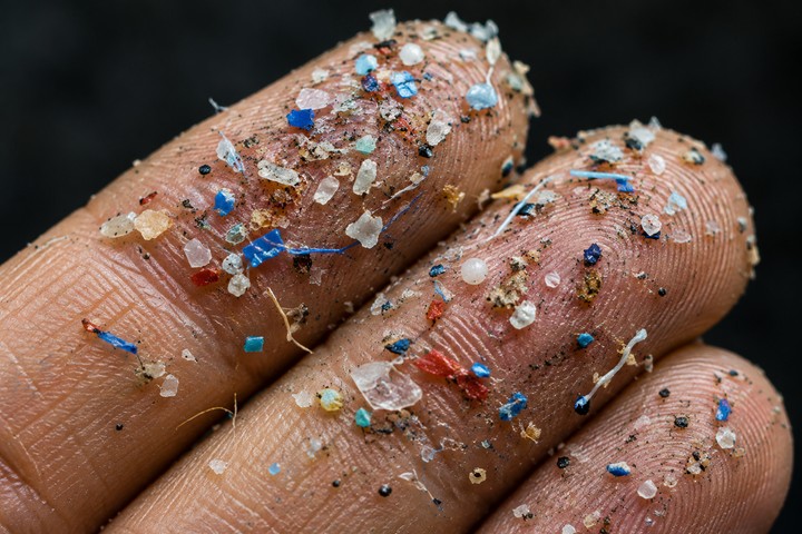
Plastics, rubber, mulch, adhesives, clothing fibers
What I feel
- Head pressure
- Brain fog
- Feeling of suffocation
- Reduced blood flow to the rest of the body
Why it looks like this
The tell: builds up over time and persists permanently after the exposure ends. Less adhesive material may migrate around the body.
My experience
Bedsheet and blanket fibers: breathing them in overnight reliably clogs my head and brings on brain fog and temperature intolerance the next day.
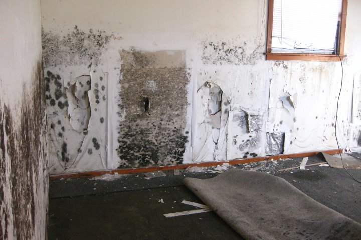
Drives stage 4 — interstitial suffocationMold
Damp buildings, outdoor spore load blowing in
What I feel
- Flu-like symptoms and heavy malaise
- Feeling feverish without running a fever
- Vascular pain — as though the arteries into my head were stiff and inflamed
Why it looks like this
The tell: effects are temporary. Of everything here, mold is the one I would choose — it can be cleaned, filtered and removed.
My experience
A mold test found heavy growth outside the building, likely blowing in through the windows, and the symptoms stopped when I wore a mask indoors.
Aggravates, does not driveMinor exposure categories
Alongside the four main categories, there are some minor ones.
Metals. Metals such as aluminum enter the air the same way the other exposures do. They impair function by inhibiting enzymatic and nervous system activity.
Oils. Oils behave like the polymers and fibers in that they hinder perfusion. Because they are fluid and amorphous, though, they don't cause the same pressure build-up, and they are easier to wash out with the right type of binder.
This pattern is not unique to me. An independent published cohort study of women with ME/CFS — not my own data or tracking — found self-reported pain and fatigue tracking measured air quality, worsening sharply once air quality moved into the "unhealthy for sensitive groups" range (see the evidence page, E21).
Four models for post-exertional malaise
PEM is usually framed as a response to exertion. In my own case, four different explanations fit the observations better (M8).
Mechanical stress
Because some symptoms track with mechanical stress, we could hypothesize an environmentally-mediated model of PEM, where particulate abrasion causes structural fragility. Because mechanical stress is strongly correlated with exertion, it would be easy to conclude the symptoms are post-exertional, but they could be post-mechanical.

Breathing rates (adult averages): U.S. EPA, Exposure Factors Handbook, 2011 edition, Chapter 6 (inhalation rates by activity level, adults 21–50). Anatomy: Servier Medical Art, CC BY 4.0.
Respiratory toxin load
Same air, same contaminant concentration, very different dose. At rest, an average adult breathes about 4–5 liters of air a minute, so only a small amount of airborne contaminant reaches the lungs and body. Increased exertion raises the respiratory rate, because the body needs to breathe more to get enough oxygen for the exertion demand. Average adult breathing rises to about 12 liters a minute during light activity, 26–28 during moderate activity and about 50 during vigorous activity, roughly 3 to 10 times the resting rate, so 3 to 10 times more airborne contaminant is inhaled and absorbed in the same period. The toxin load rises, and the result is malaise: a poisoned feeling. When patients complain about feeling poisoned, I think they are being entirely truthful, and the air they breathe is in fact poisoning them.

Waste and cell debris leave the cell into the interstitial fluid. Rising fluid pressure pushes open the overlapping endothelial flaps of the initial lymphatic (anchoring filaments hold the flaps open), and the waste and debris flow in. Downstream, a clog of plastic particles blocks the collecting lymphatic, so debris backs up and distends the vessel. Cell, organelle and red cell art: Servier Medical Art (CC BY 4.0). Lymphatic structure after Baluk et al., J Exp Med 2007.
Poor Blood Flow And Lymphatic Clearance Lead To An Exertional Susceptibility
When compressed tissue compresses the vasculature and lymphatics, less oxygen and fuel come in and less waste goes out. At rest the tissue gets by, but it has almost no energy reserve. Any extra demand drains that reserve quickly, and because drainage is clogged, metabolic waste and absorbed toxins build up faster than they can be cleared. With less oxygen and fuel arriving, cells sit at a low ATP/ADP ratio — a low energy reserve — and with lymphatic drainage blocked, waste and cell debris carried by interstitial fluid back up behind the clog, so exertion quickly drains the cell and pushes waste to harmful levels.
Autonomic neuron loss
As autonomic function is lost, the neurons that remain have to work harder to compensate. In neurons that are already overworked, normal biological activity becomes excitotoxic, and they begin to die off. Each neuron that dies leaves more work for the ones that remain, which pushes them toward the same fate and speeds up the degenerative process.
The first is mechanical stress. My pain and weakness track straining, flexing and pressure rather than muscle use. Petechiae-like dots appear on my arm after I have slept on it — load with no exertion at all. It is bad enough that I sleep with a sheet lifter, metal bars that hold the weight of the bedsheets off my body, because that weight alone is enough to make the pain and weakness worse. Exertion without strain provokes much less.
The second is respiratory toxin load. Exertion raises your respiratory rate, and a higher respiratory rate means more air, which means more airborne contaminant absorbed per minute. When I am indoors I have to hold my breathing at an almost nonexistent level to tolerate the air. Between 2019 and 2021 I made real functional gains — though nowhere close to full health — doing nothing but abdominal breathing around the clock, avoiding chest movement entirely.
What this model would explain
- Why tests come back normal. Titanium dioxide has been shown to cause Chronic Fatigue Syndrome-like behavior in mice (lethargy, passivity, loss of appetite and tremor), with minimal blood test changes despite high injection loads. The clearer damage was visible only in the tissue itself: particle deposits and lesions in the spleen and clots in the lungs' blood vessels (Chen et al., 2009). That study used high, injected doses, but the pattern is the point: the tissue showed far more than the blood.
- Why chemical sensitivity and ME/CFS overlap so heavily. Around 80% of people with ME/CFS in my surveys self-report chemical sensitivities. Given that I had no idea I had environmental issues for four years, I suspect the true figure is higher (in my opinion it’s 100%). My model of environmental drivers for symptoms explain an environmentally-triggered diagnosis.
- Why this doesn't require an acute allergic reaction. Advocating for environmental research into this disease does not mean patients need to develop hives or start wheezing when they smell perfume. Acute allergic-type reactions to transient exposure are one presentation, not a requirement — the same way lung cancer patients don't need an allergic reaction to cigarette smoke, or Parkinson's patients an allergic reaction to trichloroethylene (see the precedent above). This is a distinction I find myself having to make frequently due to the stigma that’s been placed around environmental illness (which I think developed for reasons related to chemical manufacturers and politics).
- Why the diagnostic labels blur. So frequently patients report being diagnosed with multiple conditions from the same pool including MCS, POTS, ME/CFS, Long COVID, fibromyalgia and EDS. On this model, these are versions of the same problem, differing by genetics, exposure type, and how far the disease state has progressed.
- Why brain fog and PEM might be the same mechanism. I don't think these are two separate phenomena needing two separate explanations — I think brain fog is what post-exertional malaise looks like when the interstitial suffocation described above is happening in and around the brain rather than the rest of the body.
- Why this is a histological disease, not a biopathway one. If the damage is structural — disorganized collagen fibers, clogged interstitial spaces — then biopsies and tissue imaging are what would show it, not blood work, and pharmaceuticals aimed at a pathway wouldn't be expected to fix it. That is consistent with how little blood-based and drug-trial research has found in this disease, and why the tissue imaging matters more to me than another blood panel. The findings that would amount to a high specificity biomarker are histological ones: lymphoid hyperplasia, immune infiltrates, muscle necrosis, hemosiderin deposits and microhemorrhages. Tissue depletion and barrier damage will not show up on any blood test. I believe invasive biopsies and imaging have to be done; there is no way around it.
- Why even healthy people can show some resemblances to Complex Chronic Illness. I'd guess most healthy people carry some low-grade version of this pathology and never notice, because they have nothing to compare it to and never cross the threshold into symptoms. That would make this a matter of degree, not a switch that's either on or off.
- Why this looks like a distinctly modern disease. Increased barrier permeability isn't necessarily a design flaw. Letting in more nutrients and oxygen while the barrier heals may have been a net benefit for most of human history. It only becomes a net loss once what's leaking in is more harmful than the oxygen is beneficial. Modern diet and air quality, has caused this dramatic explosion in chronic illness while genetics and infection rate remains relatively unchanged.
- Why travelers notice immediate air quality changes while visiting the United States. It isn't only the gut microbiome that shapes immune development — the environmental microbiome (the microbes naturally populating soil, forests and air) trains the immune system on what to tolerate (Kelly et al., 2022). Pollution depletes that ambient microbial population and can let more harmful strains take its place. An immune system trained on a thinner, more hostile microbial environment is a plausible second route into the same barrier-and-exposure story, independent of any single chemical.
- Why inflammatory markers differ by duration, not diagnosis. In patients who have had ME/CFS for a short time — roughly six months to two years — cytokine levels run well above healthy controls. In patients who have had it for years, cytokines run below controls: a hyperinflammatory state early on, a hypoinflammatory state later (Hornig et al., 2015). A Long COVID cohort, which by definition has not had the illness more than a few years, sits earlier on that same curve. I think most of what looks like a biomarker difference between Long COVID and ME/CFS is really a difference in how long people have been sick, not a difference in disease.
- Why mast cell treatment disappoints. Mast cell activation is downstream of barrier permeability, not upstream of it. Treating it does not address what opened the barrier.
- Why pacing works. There is a constant tug of war between healing and damage. Limiting exertion shifts the balance slightly toward healing. It does not resolve the underlying injury.
- Why a viral onset doesn't rule out an environmental driver. Titanium dioxide nanoparticle exposure has been shown to worsen respiratory syncytial virus infection in airway epithelial cells, exaggerating the same junction dysfunction the virus causes on its own. Environmental exposure and viral infection are not competing explanations for barrier damage — each can produce it, and damage from one lowers the threshold for damage from the other. That matters for anyone whose illness started with COVID or another infection and who assumes that rules out an environmental contribution.
- How this might connect to the metabolic trap hypothesis. Davis and Phair's IDO metabolic trap hypothesis proposes that ME/CFS involves a self-sustaining shift in tryptophan metabolism toward the kynurenine pathway. If gut barrier permeability increases how much tryptophan the body absorbs — plausible given the adrenergic-autoantibody research above, but not a claim either paper makes outright — that would hand the metabolic trap an upstream trigger. This is my own reading connecting two separate lines of published work, not a finding from either group.
Status of this model. This is a hypothesis developed by a patient with a research background, based on personal observation, test results, and published literature. It has not been peer-reviewed or tested in a controlled setting. The published studies cited on the evidence page are real; the synthesis and the application to ME/CFS are mine, and are unproven.
References
These are the published studies the model draws on. They are real and independent of me; the synthesis and the application to ME/CFS are my own.
- Deinhardt-Emmer S, Böttcher S, Häring C, et al. SARS-CoV-2 causes severe epithelial inflammation and barrier dysfunction. J Virol. 2021;95(10):e00110-21. ↩
- Brun E, Barreau F, Veronesi G, et al. Titanium dioxide nanoparticle impact and translocation through ex vivo, in vivo and in vitro gut epithelia. Part Fibre Toxicol. 2014;11:13. ↩
- Smallcombe CC, Harford TJ, Linfield DT, et al. Titanium dioxide nanoparticles exaggerate respiratory syncytial virus-induced airway epithelial barrier dysfunction. Am J Physiol Lung Cell Mol Physiol. 2020;319(3):L481–96. ↩ ↩
- Setyawati MI, Tay CY, Chia SL, et al. Titanium dioxide nanomaterials cause endothelial cell leakiness by disrupting the homophilic interaction of VE-cadherin. Nat Commun. 2013;4:1673. ↩
- Scheibenbogen C, Wirth KJ. Key pathophysiological role of skeletal muscle disturbance in post COVID and myalgic encephalomyelitis/chronic fatigue syndrome (ME/CFS): accumulated evidence. J Cachexia Sarcopenia Muscle. 2025;16(1):e13669. Published online December 2024. PMC11671797. ↩
- Bizjak DA, Ohmayer B, Buhl JL, et al. Functional and morphological differences of muscle mitochondria in chronic fatigue syndrome and post-COVID syndrome. Int J Mol Sci. 2024;25(3):1675. ↩
- Aschman T, Wyler E, Baum O, et al. Post-COVID exercise intolerance is associated with capillary alterations and immune dysregulations in skeletal muscles. Acta Neuropathol Commun. 2023;11:193. ↩
- Slaghekke A, et al. Microvascular dysfunction and basal membrane thickening in skeletal muscle in ME/CFS and post-COVID. Poster, International ME/CFS Conference 2025. Amsterdam UMC. ↩
- EFSA Panel on Food Additives and Flavourings. Safety assessment of titanium dioxide (E171) as a food additive. EFSA Journal. 2021;19(5):6585. ↩
- Nemat-Gorgani M, Jensen MA, Davis RW. The glymphatic–meningeal lymphatic system and autophagy in ME/CFS and Long COVID: a mechanistic perspective. Int J Mol Sci. 2026;27(20):9351. ↩
- Mitsuhashi N, Fujieda K, Tamura T, Kawamoto S, Takagi T, Okubo K. BodyParts3D: 3D structure database for anatomical concepts. Nucleic Acids Res. 2009;37(Database issue):D782–5. Model data: BodyParts3D, © The Database Center for Life Science, licensed under CC Attribution-Share Alike 2.1 Japan; the brain surface in the Model page’s transparent-brain figure is adapted from it (parts merged, interior filled, simplified). ↩
- Hickok G, Poeppel D. The cortical organization of speech processing. Nat Rev Neurosci. 2007;8(5):393–402. ↩
- Zatorre RJ, Chen JL, Penhune VB. When the brain plays music: auditory–motor interactions in music perception and production. Nat Rev Neurosci. 2007;8(7):547–558. ↩
- Koch M. The neurobiology of startle. Prog Neurobiol. 1999;59(2):107–128. ↩
- Joseph P, Arevalo C, Oliveira RKF, et al. Insights from invasive cardiopulmonary exercise testing of patients with myalgic encephalomyelitis/chronic fatigue syndrome. Chest. 2021;160(2):642–51. ↩
- Chen J, Dong X, Zhao J, Tang G. In vivo acute toxicity of titanium dioxide nanoparticles to mice after intraperitoneal injection. J Appl Toxicol. 2009;29(4):330–7. ↩
- Kelly MS, Bunyavanich S, Phipatanakul W, Lai PS. The environmental microbiome, allergic disease, and asthma. J Allergy Clin Immunol Pract. 2022;10(9):2206–17. ↩
- Hornig M, Montoya JG, Klimas NG, et al. Distinct plasma immune signatures in ME/CFS are present early in the course of illness. Sci Adv. 2015;1(1):e1400121. ↩
- Kashi AA, Davis RW, Phair RD. The IDO metabolic trap hypothesis for the etiology of ME/CFS. Diagnostics (Basel). 2019;9(3):82. ↩